Foot Muscles Mri : Peroneus Brevis Muscle Radiology Reference Article Radiopaedia Org. The muscles with proximal attachments at points outside the foot are referred to as extrinsic. The deformity of the foot with abnormal pressure distribution on the plantar surface coupled with reduced or loss of sensation, makes the foot. Head, neck, arm, foot, pelvis, etc. Abdm, abductor digiti minimi muscle; Mri patterns of neuromuscular disease involvement thigh & other muscles 2.
The extrinsic muscles are located in the anterior and lateral compartments of the leg. Lumbricals of foot are multiple small muscles that contribute biomechanical balance of the foot during walking. The flexor digiti minimi brevis (flexor brevis minimi digiti, flexor digiti quinti brevis) lies under the metatarsal bone on the little toe, and resembles one of the interossei. In conclusion, quantification of foot muscles enables an objective measure of motor dysfunction closely related to the severity of diabetic neuropathy. Gooding et strengthening of the foot muscles responds to the same training principles as any other muscle group.
Https Encrypted Tbn0 Gstatic Com Images Q Tbn And9gctvzvilsd2pu6e P62swwemme2nrmnnb11qwyhujb Fcx6yz5i Usqp Cau from The foot is a complex structure whose functions are governed by numerous muscles, ligaments, tendons, nerves and joints that work together to provide balance and stability and produce movement. Models of foot function explore different models of foot function with podiatrist kevin. Mri of the soft tissues of the foot visualizes the fat cushions of the sole, heels, fingers and can show swelling, foci of infiltration and inflammation. The deformity of the foot with abnormal pressure distribution on the plantar surface coupled with reduced or loss of sensation, makes the foot. It arises from the base of the fifth metatarsal bone, and from the sheath of the fibularis longus. The muscles of the foot can be separated into two distinct groups the extrinsic muscles of the foot arise from the anterior, posterior and lateral compartments of the leg muscles. Subscribe to foot & ankle problems. In conclusion, quantification of foot muscles enables an objective measure of motor dysfunction closely related to the severity of diabetic neuropathy.
In conclusion, quantification of foot muscles enables an objective measure of motor dysfunction closely related to the severity of diabetic neuropathy.
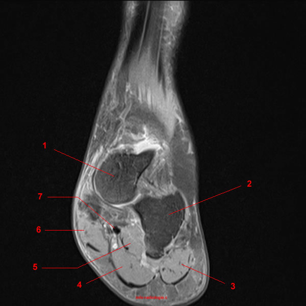
The abductor digiti minimi muscle is on the lateral side of the foot and contributes to the large lateral plantar eminence on the sole. Webmd's feet anatomy page provides a detailed image and definition of the parts of the feet and the feet are flexible structures of bones, joints, muscles, and soft tissues that let us stand upright. Learn more details about them at kenhub! Lateral and medial processes of calcaneal tuberosity. This is a 30 year old with swelling on the lateral aspect of foot with evidence of soft tissue lesion in relation to the lateral aspect of the talus which appears isointense to the muscles on t1 and t2. Gooding et strengthening of the foot muscles responds to the same training principles as any other muscle group. Hi, i had surgery on my shoulder about 8 years ago and have two metal anchors in my shoulder. The foot is a complex structure whose functions are governed by numerous muscles, ligaments, tendons, nerves and joints that work together to provide balance and stability and produce movement. Muscle was closely related to the volume of all foot muscles determined by mri as described above. Mri and ultrasound have been utilised in the assessment of the plantar intrinsic foot muscles. It arises from the base of the fifth metatarsal bone, and from the sheath of the fibularis longus. Indications for foot mri scan. The flexor digiti minimi brevis (flexor brevis minimi digiti, flexor digiti quinti brevis) lies under the metatarsal bone on the little toe, and resembles one of the interossei.
Mri findings of muscle edema or high ck levels generally differentiate a relapse of myositis from. .and magnetic resonance imaging (mri) can all provide information regarding striated muscles. Indications for foot mri scan. The foot is a complex structure whose functions are governed by numerous muscles, ligaments, tendons, nerves and joints that work together to provide balance and stability and produce movement. Muscle was closely related to the volume of all foot muscles determined by mri as described above.
Muscle Histology Vs Mri In Duchenne Muscular Dystrophy Neurology from n.neurology.org Magnetic resonance imaging (mri), with its multiplanar capabilities, superior soft tissue contrast, excellent spatial resolution, ability to image bone marrow, noninvasiveness, and lack… Models of foot function online course: In conclusion, quantification of foot muscles enables an objective measure of motor dysfunction closely related to the severity of diabetic neuropathy. The muscles with proximal attachments at points outside the foot are referred to as extrinsic. Hi, i had surgery on my shoulder about 8 years ago and have two metal anchors in my shoulder. .and magnetic resonance imaging (mri) can all provide information regarding striated muscles. A magnetic resonance imaging (mri) was performed on a normal subject; Subscribe to foot & ankle problems.
Mri with hardware in foot?
Related online courses on physioplus. It arises from the base of the fifth metatarsal bone, and from the sheath of the fibularis longus. Webmd's feet anatomy page provides a detailed image and definition of the parts of the feet and the feet are flexible structures of bones, joints, muscles, and soft tissues that let us stand upright. Mri with hardware in foot? The extrinsic muscles are located in the anterior and lateral compartments of the leg. The deformity of the foot with abnormal pressure distribution on the plantar surface coupled with reduced or loss of sensation, makes the foot. In addition, an image of all the muscles of the back and. Learn more details about them at kenhub! Mri findings of muscle edema or high ck levels generally differentiate a relapse of myositis from. Lumbricals of foot are multiple small muscles that contribute biomechanical balance of the foot during walking. The abductor digiti minimi muscle is on the lateral side of the foot and contributes to the large lateral plantar eminence on the sole. Models of foot function explore different models of foot function with podiatrist kevin. The foot is a complex structure whose functions are governed by numerous muscles, ligaments, tendons, nerves and joints that work together to provide balance and stability and produce movement.
Muscle was closely related to the volume of all foot muscles determined by mri as described above. Mri with hardware in foot? By muhammad ali, mb bs; Lumbricals of foot are multiple small muscles that contribute biomechanical balance of the foot during walking. In addition, an image of all the muscles of the back and.
Mri Of The Ankle Detailed Anatomy W Radiology from w-radiology.com Mri of the soft tissues of the foot visualizes the fat cushions of the sole, heels, fingers and can show swelling, foci of infiltration and inflammation. .magnetic resonance imaging (mri) or ultrasound imaging (usi) (soysa et al., 2012; Mri patterns of neuromuscular disease involvement thigh & other muscles 2. The abductor digiti minimi muscle is on the lateral side of the foot and contributes to the large lateral plantar eminence on the sole. Models of foot function online course: Subscribe to foot & ankle problems. Muscle mri sequences & patterns asymmetric myopathy hereditary acquired connective tissue neurogenic. .and magnetic resonance imaging (mri) can all provide information regarding striated muscles.
Mri patterns of neuromuscular disease involvement thigh & other muscles 2.
Models of foot function explore different models of foot function with podiatrist kevin. Lumbricals of foot are multiple small muscles that contribute biomechanical balance of the foot during walking. Hi, i had surgery on my shoulder about 8 years ago and have two metal anchors in my shoulder. Feet and ankles ankle muscle anatomy of foot muscles of foot muscles foot foot muscles anatomy muscle composite video showing multiple mri images including: Mri findings of muscle edema or high ck levels generally differentiate a relapse of myositis from. Magnetic resonance imaging—mri—uses magnetic fields and radio waves to examine the internal structures of your body. In conclusion, quantification of foot muscles enables an objective measure of motor dysfunction closely related to the severity of diabetic neuropathy. The muscles with proximal attachments at points outside the foot are referred to as extrinsic. The muscles of the foot can be separated into two distinct groups the extrinsic muscles of the foot arise from the anterior, posterior and lateral compartments of the leg muscles. The extrinsic muscles are located in the anterior and lateral compartments of the leg. Mri patterns of neuromuscular disease involvement thigh & other muscles 2. Related online courses on physioplus. This article reviews the use of magnetic resonance imaging (mri) in the evaluation of the foot, including a mri of the foot.
Share :
Post a Comment
for "Foot Muscles Mri : Peroneus Brevis Muscle Radiology Reference Article Radiopaedia Org"


Post a Comment for "Foot Muscles Mri : Peroneus Brevis Muscle Radiology Reference Article Radiopaedia Org"